-
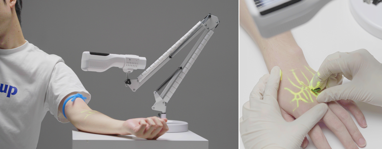
How can vein finder complement the workflow of ophthalmic medicine and imaging equipment?
Nov 27 , 2025
1) Core Value: Provide a unique "surface vascular map" Ophthalmic imaging equipment (such as OCT) focuses on the microscopic vascular structure of the fundus (retina, choroid), while venous detectors are adept at non-invasively visualizing the vascular network on the ocular surface and around the eye, especially: The blood vessels of the sclera and conjunctiva The blood vessels of the skin around ...
Read More
-
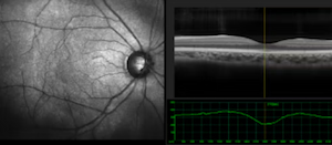
Early detection of diabetic retinopathy using OCT imaging
Jan 22 , 2026
1) Traditional Screening Methods and Their Limitations Traditionally, the screening and staging of DR Mainly relied on color fundus photography to observe two-dimensional surface lesions such as retinal microaneurysms, hemorrhage and exudation. However, this method has limitations: Strong subjectivity: Relying on the doctor's experience. Inability to observe deep structures: It is impossible to qu...
Read More
-
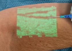
How deep can a medical vein finder see?
Jan 27 , 2026
1. Main detection depth General depth: Most devices can detect veins 0.2 to 1.0cm beneath the skin, but the actual effective imaging range is usually concentrated on the shallower veins under the skin (0.2 to 1.0 cm). Best effect: For clearly visible superficial veins (such as the back of the hand and forearm), the imaging effect is the best. 2. Key influencing factors Skin condition Skin color, f...
Read More
-
The clinical application of venous imaging equipment in oncology departments and chemotherapy wards
Apr 03 , 2026
The main value of the venous imaging device in oncology departments and chemotherapy wards lies in addressing the "difficult needle insertion" problem faced by chemotherapy patients due to repeated punctures and deteriorating vascular conditions. It not only significantly improves the success rate of the first puncture, shortens the operation time, but also reduces the pain experience of patients ...
Read More
-
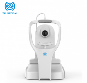
Can one drive after having the OCT eye examination?
May 06 , 2026
1. If it is a "non-mydriatic" examination (without dilating the pupils) - one can drive directly Many of the current high-end OCT devices (especially in routine screenings) have the feature of non-mydriatic examination. If the patient did not receive any eye drops to dilate the pupils before the examination, but simply looked at the cursor of the instrument for a while: Visual status: After the ex...
Read More


 IPv6 network supported
 English
 español
 العربية


