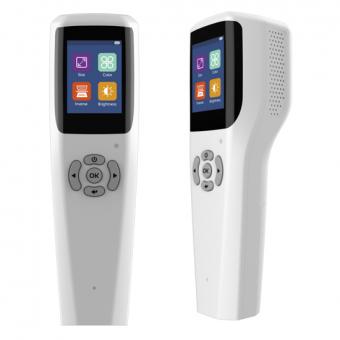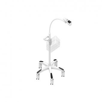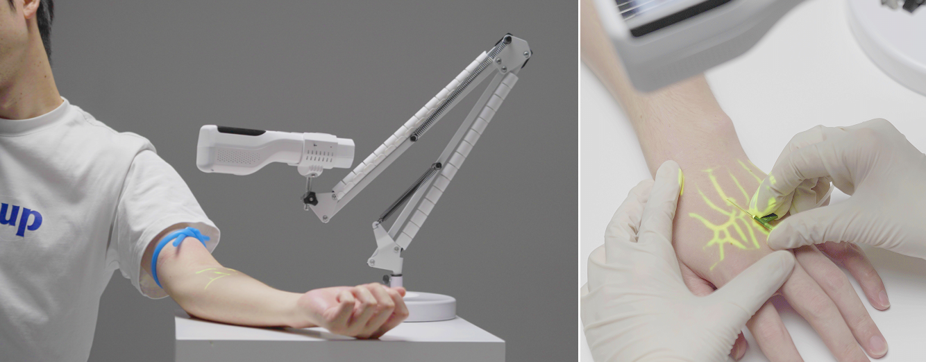

Every year on National Eye Care Day, it is a warm initiative to promote the overall eye health of the entire population. This year, we focus on the common vision of "(Vision for Everyone)" - this is not just a slogan, but also a profound pursuit of accessibility to eye health services. Often, the "alarms" of the eyes remain silent. Blurred vision, dryness and fatigue could be signs of excessive eye use or early symptoms of underlying eye diseases. Precise examinations are the first step to dispel the fog and achieve effective management. As an enterprise deeply involved in the field of ophthalmic diagnostic equipment, Zhiding Medical is committed to ophthalmic devices such as optical coherence tomography (OCT) and dry eye detectors. It integrates AI and remote diagnosis to break through geographical and resource barriers, and helps turn the vision of "ensuring everyone has eye health" from a vision into reality. It's not just "dry eyes": Let dry eye diagnosis and treatment bid farewell to "judging by feelings" "Dry eyes, a foreign body sensation, photophobia..." These are the most common eye discomforts, often simply attributed to fatigue. However, true "dry eye" is a multi-factorial eye surface disease with complex types. Blind treatment may have the opposite effect. The dry eye diagnosis and treatment plan of Zhiding Medical begins with precise assessment: not only focusing on symptoms, but also through professional tests such as tear film quality analysis and meibomian gland imaging, it clearly reveals the "root cause" of dry eye - whether it is "lack of moisture" (insufficient tear secretion), "lack of oil" (meibomian gland dysfunction), or a combination of both. By clearly identifying the type and formulating individualized treatment plans, it can address the discomfort at its source, improve visual quality and enhance life comfort. Fundus OCT examination: Insights into the fundus, preventing problems before they occur If ocular surface problems cause discomfort, then the lesions in the fundus of the eye may quietly steal vision. Hypertension, diabetes, high myopia, or as one ages, all can cause problems in the "life support system" of the eye such as the retina, macula, and optic nerve. Optical coherence tomography (OCT) is hailed as the "CT" of ophthalmology. It does not require contact with the eyeball and can obtain high-resolution tomographic images of the fundus tissue within seconds, with an accuracy of micrometers. It is a non-invasive and high-resolution eye examination technique. Why is it important? Many eye diseases are difficult to detect during routine vision examinations, but OCT can achieve this painlessly: Early screening: Before symptoms appear, timely detection of optic nerve damage, macular edema, holes, etc. in glaucoma. Precise diagnosis: Clearly distinguish the structures of each layer of the retina, providing key evidence for the diagnosis of macular degeneration, diabetic retinopathy, retinal vein occlusion, etc. Eff...
Read More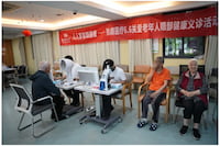
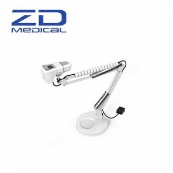
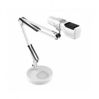
On June 6th, which is the National Eye Care Day, with the noble mission of "ensuring everyone enjoys good eye health", ZD Medical broke the barrier of medical access distance and brought professional eye care equipment to a nursing home in Hangzhou to conduct a free public eye care consultation activity. They delivered professional eye health public welfare eye care services to the elderly. Dry eyes, frequent eye rubbing, eye mites, and blurred vision are common daily troubles for many elderly people in the nursing home. Many elders suffer from eye discomfort but rarely have the opportunity for professional eye examinations. This free medical consultation event is open to all senior citizens in the hospital. Professional equipment such as dry eye detection instruments, ophthalmic microscopic image analysis equipment, and eye misting devices are fully equipped. The event provides a one-stop service offering multiple free eye examinations and health education. Staff members carefully examined the tear secretion of the attending seniors, checked for meibomian gland mites, and accurately screened potential risks of dry eye syndrome. On-site, the seniors had the opportunity to experience eye misting inhalation therapy for quick relief of dry eyes, eye fatigue, and foreign body sensation. At the event, staff members provided one-on-one interpretation of the eye examination reports, combined with the seniors' physical conditions, to explain the prevention knowledge of common eye diseases such as cataracts and dry eye syndrome. They also customized home eye care tips and gave all the participants in the screening a heartwarming care gift fromZD Medical. This public welfare initiative representsZD Medicalcommitment to fulfilling its social responsibilities and its dedicated efforts in promoting eye health for all. Leveraging its own equipment development advantages,ZD Medicalhas consistently adhered to the principle of providing convenient public welfare services to safeguard the eye health of the entire population, and has been practicing its social responsibilities through professional ophthalmic techniques. In the future, we will continue to expand our public welfare resources and regularly conduct free medical consultations, so that professional eye health services can benefit more elderly people across the country. Also welcome tocontact us, we are ZD Medical Inc. Tel : +86-187 9586 9515 Email :sales@zd-med.com Whatsapp/Mobile : +86-187 9586 9515
Read More
Also welcome to contact us, we are ZD Medical Inc. Tel : +86-187 9586 9515 Email :sales@zd-med.com Whatsapp/Mobile : +86-187 9586 9515
Read More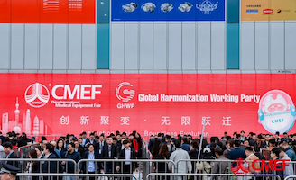
Spring tides surge, innovation leads the way. From April 9th to 12th, 2026, the 93rd China International Medical Equipment Fair (CMEF) concluded successfully at the Shanghai National Exhibition Center. As a global benchmark for the medical and health industry, this exhibition, with the theme of "Innovation Fusion - Infinite Transformation", brought together nearly 5,000 brands from around the world and over 120,000 professional visitors, creating a trading and technical exchange platform covering the entire industry chain and linking global resources. ZD Medical showcased its core products such as the OCT series, dry eye diagnosis equipment, and vascular imaging devices at the G57 booth in Hall 7.2. With its meticulous dedication and innovative spirit in the field of ophthalmology, it collaborated with global industry peers to explore the frontiers and discuss development, thereby injecting Chigeng's strength into this grand event. 1 - As a high-tech enterprise specializing in ophthalmology, ZD Medical has been deeply engaged in the research, production and sales of high-precision optical medical equipment in ophthalmology since its establishment in 2015. With over 90 patent authorizations, it has become a benchmark force for independent innovation in the industry. At this CMEF exhibition, the product portfolio of Zhiding Medical became the center of attention, with a continuous flow of visitors. The on-site staff patiently explained the product principles, operation procedures and core advantages, and meticulously answered various questions. The professional service and high-quality products won unanimous recognition and praise from the guests present, making ZD Medical one of the most popular booths in Hall 7.2. 2 - At this exhibition, ZD Medical fully showcased its innovative achievements in the field of ophthalmic diagnosis, covering areas such as fundus diagnosis, dry eye treatment, and vascular imaging, presenting a comprehensive brand strength of "research, production, and sales" in all aspects. It provided the industry with an eye care comprehensive solution that is both professional and cost-effective, and became the core highlight of the exhibition. From the all-process intelligent diagnostic OCT series to the highly efficient and precise dry eye diagnosis equipment, and then to the highly practical vascular imaging instrument, each product has attracted the attention of numerous ophthalmologists, industry partners and professional visitors with its outstanding technical advantages and efficient clinical application value. They all came to stop and watch, and queue up to experience it. 3-A Successful Conclusion: Keeping the Original Intent, Reaching for Light and Starting a New Journey The curtain of the 93rd CMEF has fallen, but ZD Medical's exploration journey in the field of ophthalmic diagnosis and treatment has never stopped. This CMEF exhibition is not only a showcase window for brand innovation achievements, but also an important...
Read More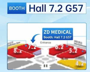
Spring awakens all things, bright eyes create new chapters. The 93rd China International Medical Equipment Fair (CMEF) will grandly open at the Shanghai National Exhibition Center from April 9th to 12th, 2026. At that time, ZD Medical will showcase its high-quality products such as OCT, dry eye diagnosis equipment, and vascular imaging devices. We sincerely invite you to visit the G57 booth in Hall 7. Let's explore the cutting-edge innovations together and jointly build a new future for the healthcare industry. Also welcome to contact us, we are ZD Medical Inc. Tel : +86-187 9586 9515 Email : sales@zd-med.com Whatsapp/Mobile : +86-187 9586 9515
Read More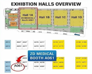
Time: March 30th - April 2nd, 2026 Location: Hangzhou International Expo Center (HIEC) Highlights: Fundus & Dry Eye series new product launch, on-site experience of top-notch ophthalmic black technology. ZD Medical has always adhered to the principle of "protecting vision with technology". On the occasion of the 38th Zhejiang International Scientific Research Exhibition, we will meet you in Hangzhou with a sense of awe for life and an ultimate pursuit of technology. From fundus imaging to dry eye analysis, every piece of equipment embodies our thoughts on precision medicine. Step into booth A061 and feel the warmth of medical technology. Also welcome to contact us, we are ZD Medical Inc. Tel : +86-187 9586 9515 Email : sales@zd-med.com Whatsapp/Mobile : +86-187 9586 9515
Read More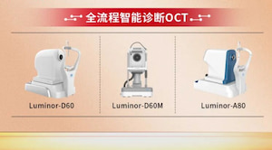
Body Text: ZD Medical is proud to announce that our proprietary Optical Coherence Tomography (OCT) system has successfully been shortlisted in the bidding process for the 2026 Spring Zhejiang Medical Equipment Exhibition. This selection marks a significant milestone in our market expansion strategy and serves as a testament to the clinical precision and technological innovation of our ophthalmic solutions. The "Zhejiang Medical Exhibition" procurement cycle is one of the most rigorous evaluation processes in the region, assessing medical devices based on strict criteria regarding performance, quality control, and cost-effectiveness. The successful inclusion of ZD Medical OCT system demonstrates our product's ability to compete with top-tier international brands and meet the high standards required by public hospitals and medical institutions. "Being selected for this procurement catalog is not just a commercial win; it is a validation of our R&D commitment," at ZD Medical. "It confirms that our OCT technology provides the high-definition imaging and reliability that clinicians demand, positioning us strongly for broader adoption in the domestic and international markets." As ZD Medical continues to grow, we remain dedicated to providing cutting-edge ophthalmic diagnostic solutions to practitioners worldwide.
Read More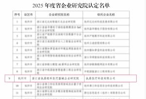
At the beginning of the New Year, when everything takes on a fresh look. After strict review and recognition by the Zhejiang Provincial Department of Economy and Information Technology, ZD Medical has been officially approved to establish the "Zhejiang ZD Medical Ophthalmic Medical Device Enterprise Research Institute". This is a high recognition of the company's continuous dedication to the high-end medical device field for ophthalmology, its commitment to technological innovation and R&D investment. It also marks a crucial step taken by ZD Medical in building a high-level R&D system and leading the technological development of the industry. As a technology-driven enterprise, ZD Medical has always been guided by clinical needs and has been deeply engaged in the field of ophthalmic medical innovation. The recognition of this provincial enterprise research institute marks that ZD Medical has officially entered the "first echelon" of medical device innovation in the province, and will build a higher-level platform for the construction of a collaborative innovation system integrating "industry, academia, research and medicine". As the New Year begins, the mission is on our shoulders. In the future, ZD Medical will take the provincial enterprise research institute as the core engine, continuously increase investment in research and development, focus on the breakthroughs and transformation of core ophthalmic technologies, and empower clinical practice and safeguard the eye health of the general public with higher-quality medical products. Thank you to every partner for your side by side. In the New Year, ZD Medical will take this honor as a new starting point, keep moving forward on the road of innovation, and inject stronger impetus into the high-quality development of the ophthalmic medical industry!
Read More

 IPv6 network supported
 English
 español
 العربية