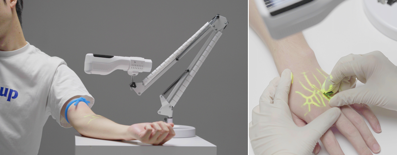
1) Core Value: Provide a unique "surface vascular map"
Ophthalmic imaging equipment (such as OCT) focuses on the microscopic vascular structure of the fundus (retina, choroid), while venous detectors are adept at non-invasively visualizing the vascular network on the ocular surface and around the eye, especially:
The blood vessels of the sclera and conjunctiva
The blood vessels of the skin around the eyes and the eyelid margin
This provides ophthalmologists with a brand-new dimension of observation.
2) Integration and supplementation of specific work processes
1. Diagnosis and Screening stage: Viewing "Systemic Diseases" from "Ocular Surface Blood Vessels"
Many systemic diseases can affect both the fundus blood vessels and the ocular surface blood vessels simultaneously.
Venous detection devices can serve as a rapid and non-invasive initial screening tool.
Supplement the workflow of OCT
Scene: A diabetic patient comes for an annual eye examination.
The current process: The doctor uses OCT to examine the retinal microvessels, looking for signs of diabetic retinopathy such as microaneurysms and non-perfusion areas.
Supplementary value: At the initial stage of the consultation, a venous detector is first used to observe the blood vessels of the patient's bulbar conjunctiva. Research has found that systemic diseases such as diabetes can cause morphological changes such as distortion and dilation of conjunctival blood vessels. If any abnormalities are detected, it can provide stronger directivity for
OCT examination and alert doctors that the patient may have uncontrolled systemic risks.
Advantages: The examination process is faster and more comfortable, with high patient tolerance, and it is easy to conduct dynamic observation.
2. Surgical planning and navigation: Enhance the precision and safety of the surgery
This is one of the most promising application scenarios of venous detectors in ophthalmology.
Supplement the workflow of cataract surgery
For cataract patients with limbal stem cell insufficiency, thin sclera or those requiring complex incisions, it is crucial to understand the vascular distribution in the limbal area of the corneosclera and the surgical incision area before the operation. This can prevent intraoperative bleeding and affect the surgical field of view.
Supplementary value: Before the operation, a venous detector is used to draw the vascular network of the corneoscleral margin, which helps doctors precisely plan the position of the surgical incision, avoid the main blood vessels, achieve "blood-free" surgery, and improve the efficiency and safety of the operation.
Supplement the workflow of glaucoma surgery
Question: When performing glaucoma drainage device implantation (such as Ah valve implantation), it is necessary to suture and fix it in the water tank area (usually located behind the equator of the eyeball). This area is rich in blood vessels, and intraoperative bleeding is a common complication.
Supplementary value: Before the operation, a venous detector is used to precisely locate the surface blood vessels in the water tank area. With the assistance of the surgical navigation system, doctors can select areas with relatively sparse blood vessels for suturing, significantly reducing the risk of bleeding and postoperative complications.
Supplement the workflow of eye plastic surgery/orbital surgery
Blepharoplasty, orbital tumor resection, etc., require an accurate understanding of the vascular anatomy of the eyelid and orbit.
Supplementary value: The venous detector can clearly outline the direction of the arteries and veins around the eyes, serving as a "real-time vascular map" for surgical navigation. It helps doctors perform precise operations, avoid damaging important blood vessels, and reduce intraoperative bleeding and postoperative hematoma.
3. Treatment and efficacy evaluation stage: Achieve visual efficacy monitoring
Supplement the dry eye treatment workflow
Meibomian gland dysfunction is the main cause of dry eye. Intense pulsed light (IPL) is an effective treatment method, and one of its mechanisms of action is to close the abnormal blood vessels in the eyelid.
Supplementary value
Before treatment: Use a venous detector to assess the vascular density and morphology of the eyelid margin to provide baseline data for treatment.
After treatment: Re-imaging is conducted to visually display the closure of abnormal blood vessels after IPL treatment, providing objective imaging evidence for efficacy evaluation. This is unique information that existing corneal topography or dry eye analyzers cannot provide.
Supplement the treatment workflow for ocular surface inflammatory diseases:
Allergic conjunctivitis, pterygium and other diseases are accompanied by obvious conjunctival vascular congestion.
Supplementary value: Quantitative analysis of congested blood vessels (such as vessel diameter and density) using a venous detector can objectively evaluate the effects before and after anti-inflammatory drug treatment, providing data support for clinical research and personalized treatment.
Also welcome to contact us, we are ZD Medical Inc.
Tel : +86-187 9586 9515
Email : sales@zd-med.com
Whatsapp/Mobile : +86-187 9586 9515


 IPv6 network supported
 English
 español
 العربية


