-
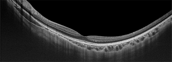
Have you got the OCT image of the fundus, are you still worrying about how to interpret it?
Dec 20 , 2024
Welcome to science time! This article will discuss optical coherence tomography (OCT), an advanced technology widely used in eye examination, and introduce the basic principles and sequence of OCT image reading to help people better understand this convenient and non-invasive ophthalmic examination tool. Have you got the OCT image of the fundus, are you still worrying about how to interpret it? Lo...
Read More
-
What are the working principles and characteristics of Optical Coherence Tomography?
Jan 03 , 2025
Angio OCT (Optical Coherence Tomography) is a non-invasive imaging technique used to observe the vascular structure and blood flow dynamics of the retina and choroid at high resolution. It combines the cross-sectional imaging capabilities of optical coherence tomography (OCT) with blood flow analysis and is widely used in the field of ophthalmology. Working principle The core technology of Angio O...
Read More
-
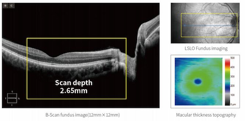
What are the features and benefits of Optical Coherence Tomography Luminor-D60?
Jan 11 , 2025
ZD Medical new generation latest upgraded version of split OCT + anterior eye segment function Model: Optical Coherence Tomography Luminor-D60 LSO technology is used for fundus imaging, which accurately helps you to identify retinal diseases, helps screening and reduce the leakage of diseases in the initial examination, and can greatly improve the efficiency of clinical use. The ZD...
Read More
-
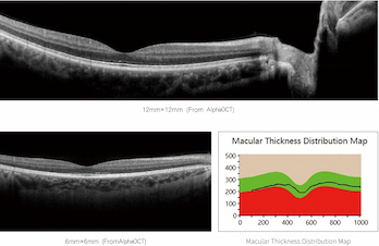
What does optical coherence tomography test for?
Jan 11 , 2025
Optical Coherence Tomography (OCT) is a non-invasive imaging test that provides high-resolution cross-sectional images of tissues. It is widely used in ophthalmology and other medical fields to assess various conditions. Here's what it typically In Ophthalmology OCT is most commonly used to evaluate structures within the eye, particularly the retina and optic nerve, for the following c...
Read More
-
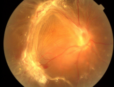
What causes retinal detachment?
Mar 13 , 2025
Retinal Detachment (RD) refers to the separation of retinal nerve epithelia from pigment epithelia, resulting in the inability of photoreceptor cells to properly receive and conduct light signals, which can lead to irreversible visual impairment or even blindness in severe cases. RD can be divided into three categories: porosity, tractive and exudative. Among them, rheogenic retinal detachment is ...
Read More
-
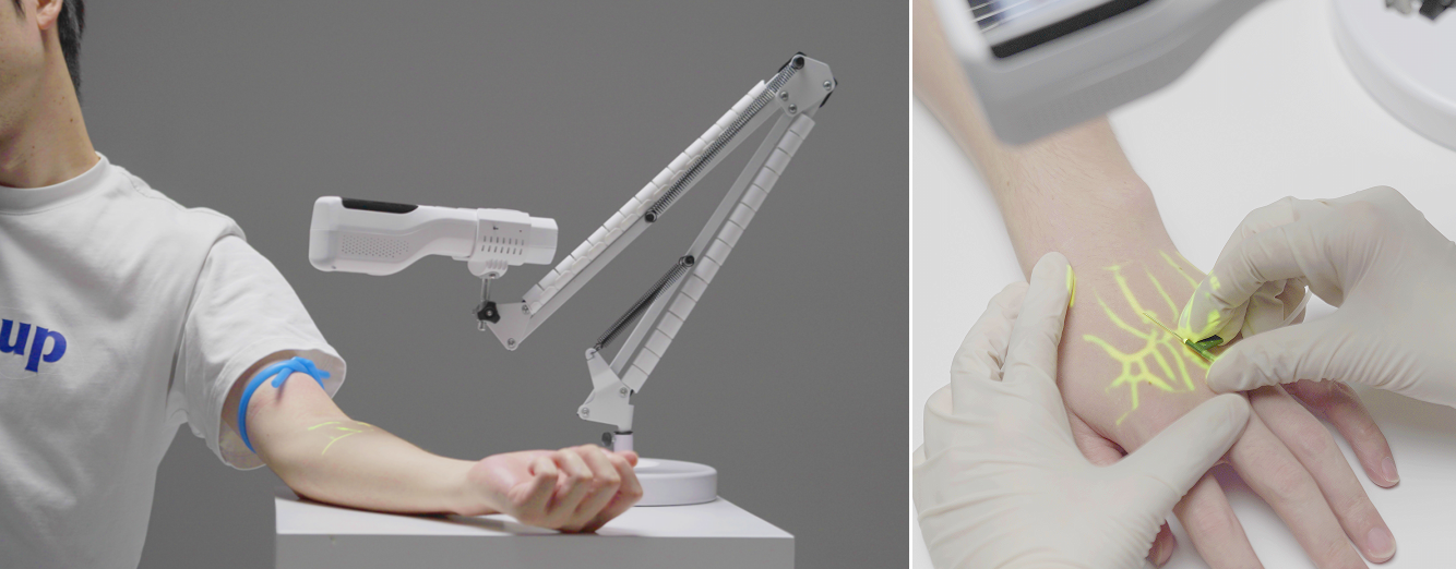
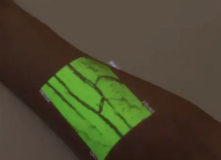
What are the benefits of using a Vein finder?
Mar 21 , 2025
As a medical aid device, Vein Finder has significant advantages in clinical operation, especially in Settings such as venipunction and vascular evaluation, which improves efficiency and patient experience. Here is a comprehensive analysis of its core strengths: 1. Improve clinical operation efficiency and success rate - High first-time puncture success rate: Through near-infrared light technology,...
Read More
-
What color light helps find veins?
Mar 27 , 2025
In medical practice, green light (with a wavelength of about 540nm) is generally considered the color of visible light that is most helpful in locating veins. Here's the explanation: 1. Optical principle: - Deoxyhemoglobin in Vein Finder has a higher absorption rate of green light, while surrounding tissues (such as skin and fat) reflect more green light. This contrast between absorption and refle...
Read More
-
Is optical coherence tomography worth it?
Apr 08 , 2025
Optical coherence tomography (OCT) is a valuable imaging technology with diverse applications in medicine, particularly in ophthalmology and cardiology. Its utility depends on the clinical context, but here’s a comprehensive evaluation of its benefits and considerations: Key Benefits of OCT 1. Non-Invasive and Safe - OCT uses light waves (e.g., near-infrared) instead of radiation or invasive probe...
Read More
-
Holding hands and looking at the eyes to help with diagnosis | Suddenly
May 14 , 2025
Common causes: tractional retinal detachment (TRD) is mainly caused by the traction of the fibrous vascular membrane on the surface of the retina or within the vitreous. Adult TRD is most commonly seen in proliferative diabetic retinopathy (PDR), retinal vein occlusion (RVO), retinal vasculitis and proliferative vitreoretinopathy (PVR). In addition, trauma, sickle cell retinopathy and uveitis, etc...
Read More


 IPv6 network supported
 English
 español
 العربية


