When peripheral IV access becomes difficult, the instinct is to reach for the most powerful tool available. That instinct is understandable, and it is also the reason ultrasound machines are sometimes tied up on a 26-gauge paediatric stick while a deeper line waits in the corridor.
The real question is not "which device is better?" It is "which device is right for this patient, at this step in the pathway?"
Light-based near-infrared (NIR) vein visualization and ultrasound-guided cannulation do not compete. They occupy different tiers of a graduated access pathway — and using them in the right order is what produces the throughput and first-stick gains that get reported in the literature.
In one line: NIR visualization is the fast, zero-contact first look at superficial vessels; ultrasound is the escalation tool for depth, guidance and central access.
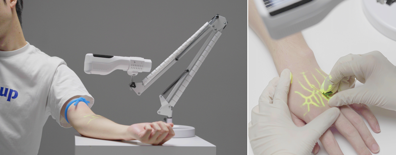
Haemoglobin absorbs near-infrared light more strongly than surrounding tissue. A vein finder illuminates the skin, captures the backscattered signal with a camera, processes the contrast digitally, and projects a vascular map back onto the skin — in real time, in the position where the vein actually is. Grown from ZD Medical's first vein visualization products in 2012, these devices are available in handheld, table-stand, flexible-stand and mobile-trolley configurations.
Ultrasound uses high-frequency sound to image vessels — and surrounding anatomy — in real time, including at depth. It is the definitive tool for deep vessels, for vessels that cannot be seen or palpated, and for central venous access. It provides dynamic needle-tip guidance, which is precisely what makes it indispensable for the hardest sticks.
The trade-off is equally clear: it requires a trained operator, a machine that may be shared across departments, a sterile probe cover and gel for each use, and a workflow that takes longer per attempt than a projection device.
|
Dimension |
Near-infrared vein finder |
Ultrasound guidance |
|
Vessel depth |
Superficial peripheral veins |
Deep and superficial, full anatomy |
|
Real-time needle guidance |
No — pre-puncture mapping |
Yes — dynamic tip tracking |
|
Operator training burden |
Low; usable at the bedside |
High; requires credentialed skill |
|
Per-attempt consumables |
Minimal |
Probe cover, gel, sterile field |
|
Equipment mobility |
Handheld or trolley; single-purpose device |
Shared, higher-cost platform |
|
Throughput for routine sticks |
High — seconds to assess, no setup |
Lower — setup per attempt |
|
Best-fit scenario |
Most superficial peripheral attempts, including difficult ones |
Deep, non-palpable, or central access |
|
Where it fits |
First-line assessment |
Escalation and rescue |
Read the table as a sequence, not a scorecard. Each row describes a division of labour.
Emergency department. High volume, heterogeneous patients, minimal time. A projection vein finder that works in ambient light and can be deployed in seconds at the bedside fits the assessment step; ultrasound is reserved for the genuinely refractory case. Confirm the device performs under bright triage lighting — not in a dimmed showroom.
Paediatrics and neonates. Small veins, limited surface area, high emotional stakes. Multiple size and depth modes — including dedicated paediatric settings — and non-contact operation matter more here than raw magnification. Fewer attempts also means less procedural distress, which has value beyond efficiency.
Oncology and infusion centres. Repeated access in patients with vascular damage from prior therapy. NIR visualization supports identifying remaining usable vessels; ultrasound supports the deeper or more compromised ones and is the appropriate tool when prior access has exhausted superficial options.
Radiology and critical care. Central and deep access is ultrasound's domain, and this is where it should stay. NIR visualization remains useful for the peripheral lines that keep the patient moving while the deeper access is planned.
Blood draw stations and outpatient phlebotomy. High-throughput, mostly superficial, often fast-turnaround. This is a form-factor question: table-stand or flexible-stand configurations suit a fixed draw station better than a handheld, and buying the wrong form factor is the most common reason a device ends up in a drawer.
Veterinary clinics and animal laboratories. The underlying technical principle is not species-specific. NIR vein visualization has found real use for venous access in pets and other mammals where veins are hard to see through fur and pigmented skin.
ZD Medical's vein finder line covers the form factors a facility actually needs — which is the practical answer to "one device cannot serve every department":
Match the configuration to the workflow first. Optics second.
Used as a last resort. The gain comes from early assessment. Fix: make it a step in the SOP, not a rescue measure.
Bought without testing skin tones and ambient light. Contrast performance across pigmentation is the single biggest differentiator between a demo unit and a clinical one. Fix: test on the darkest and lightest skin available, under real department lighting.
Projection misalignment tolerated. A few millimetres of offset is worse than no device, because it creates false confidence. Fix: verify alignment against successful cannulation during evaluation.
No owner. Devices that belong to nobody stop working within a quarter. Fix: assign charging, cleaning and monthly checks to a named role.
Not escalated when it should be. A vein finder that is used too long on a case that needed ultrasound delays care. Fix: write the escalation triggers into policy, not into individual judgement.
Is a vein finder better than ultrasound for IV access? Neither is universally better. A near-infrared vein finder is faster, lower-cost and needs minimal training for superficial peripheral veins. Ultrasound is essential for deep, non-palpable or central access because it provides real-time needle guidance. They work best in sequence.
When should you escalate from a vein finder to ultrasound? When the target vessel is not visible or palpable despite visualization, when a deep or central vessel is required, or after two failed attempts. That last trigger should be written into policy.
Does a vein finder replace clinical judgment? No. It shows where a superficial vein is; it does not assess whether that vein is suitable for the intended therapy, infusion rate or dwell time. That decision remains with the clinician.
Can a vein finder be used on patients with dark skin tones? It can, but contrast performance varies between devices. Test any candidate device on the darkest and lightest skin tones available during evaluation, not on a healthy volunteer's forearm.
Is NIR vein visualization safe? Yes. It is non-invasive, uses near-infrared light, involves no ionizing radiation, no contrast agent and no contact with the skin.
Can vein finders be used in veterinary practice? Yes. The technical principle applies to mammals generally, and NIR vein finders are already used in overseas veterinary clinics, pet hospitals and animal laboratories.
Does a vein finder work in bright environments like emergency vehicles?
Visibility depends on ambient light. Performance in emergency vehicles, operating theatres and sunlit triage areas should be verified under real department lighting before purchase.
The mature access program does not choose between light and sound. It sequences them: visualize first with near-infrared, escalate to ultrasound when indicated. Facilities that adopt that order tend to report shorter procedure times, fewer repeat attempts, and a measurable lift in first-stick success — and they stop tying up their ultrasound machine on sticks a projection device could have handled in seconds.
ZD Medical Inc. developed its first vein visualization products in 2012 and has shipped infrared vein finders in handheld, table-stand, flexible-stand and mobile-trolley configurations since. Every unit is built around the same patented near-infrared projection principle described above, and is supported by clinical onboarding for distributors and hospital teams.
Interested in becoming a distributor? Contact us for wholesale pricing.
Tell us your department mix and monthly puncture volume, and we will help you size the right configuration — and build the ROI case to go with it.
Tel / WhatsApp: +86-187 9586 9515 · Email: sales@zd-med.com


 IPv6 network supported
 English
 español
 العربية


