I. Core Technical Principles
Near-infrared imaging (NIR Technology)
The absorption characteristics of hemoglobin: Deoxyhemoglobin in veins has a higher absorption rate for near-infrared light of specific wavelengths (typically 700-900nm) than that in surrounding tissues.
Reflection difference imaging: The device emits near-infrared light onto the skin, and the camera captures the reflection signal. Through algorithms, the veins are contrasted and enhanced with the surrounding tissues to form a clear image.
Multispectral imaging (optional technology)
Some high-end devices combine visible light and near-infrared light and adapt to different skin tones (such as patients with dark skin) through multispectral analysis.
AI image processing
Edge enhancement algorithm: Highlighting vein contours and reducing noise interference.
Depth prediction: Estimate the depth of veins through reflection intensity to assist in locating deep veins.
 Ii. Enhanced Features of Desktop Design
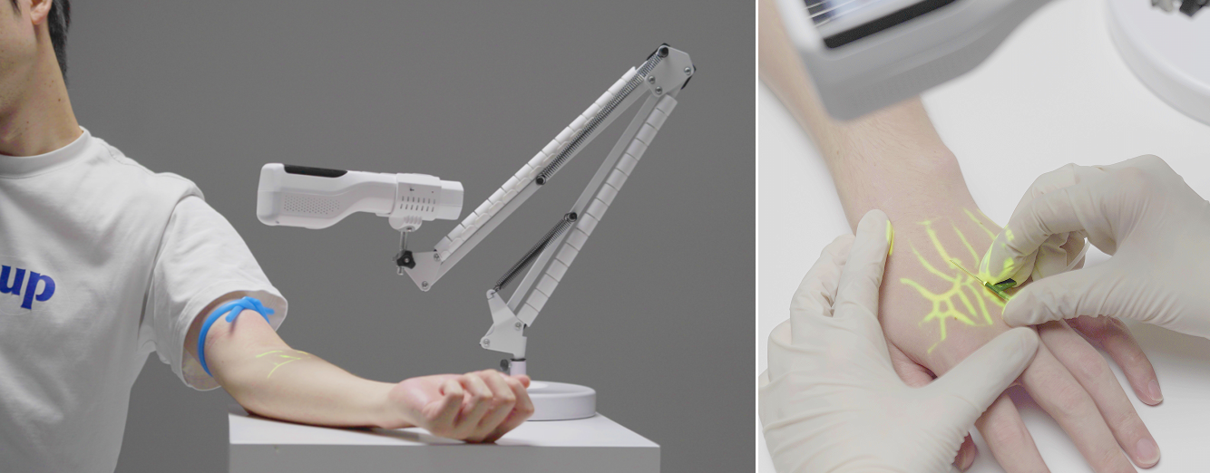
Stable projection system
Fixed projector: Avoid the shaking of
handheld devices Vein Finder and precisely project vein images onto the skin surface (such as green/red contours).
Adjustable stand: Freely adjust the height and Angle to fit different parts such as the arm, back of the hand, and foot.
High-precision camera
Equipped with high-resolution sensors, it captures subtle vascular signals in real time and, in combination with the optical zoom function, magnifies local areas.
Environmental adaptability
Automatic calibration: Dynamically optimize imaging parameters based on ambient light and skin color.
Anti-interference design: Reduce the influence of surgical lamps or other light sources on imaging.
Iii. Work Process (Taking Blood Drawing as an Example)
Step 1: Patient localization
The patient placed his arm flat under the device and kept it stable.
Step 2: Quick scan
The device emits near-infrared light, generating a vein distribution map within 1-2 seconds and projecting it in real time.
Step 3: Vascular selection
Medical staff select the best puncture site (such as thick, straight, and unbifurcated veins) through screen or projection markers.
Step 4: Precise puncture
Puncture is completed under the guidance of venous projection, reducing the number of blind explorations.
Also welcome to contact us, we are ZD Medical Inc.
Tel : +86-187 9586 9515
Email : sales@zd-med.com
Whatsapp/Mobile : +86-187 9586 9515


 IPv6 network supported
 English
 español
 العربية


